


3D Printed Osteochondroma
Clinical History
A 61-year old male with prostate cancer attends pre-assessment clinic prior to
a prostatectomy. Overall, he feels well with no major complaints. On review of symptoms, it is noted he has chronic
pain in his right knee, which his GP called osteoarthritis. To exclude boney metastases of the prostate carcinoma, a
knee x-ray is ordered, which shows a pedunculated lesion projecting from the medial aspect of the diaphysis of the
right femur. His prostatectomy goes ahead but he subsequently dies from a postoperative pulmonary embolism.
Pathology
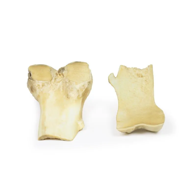
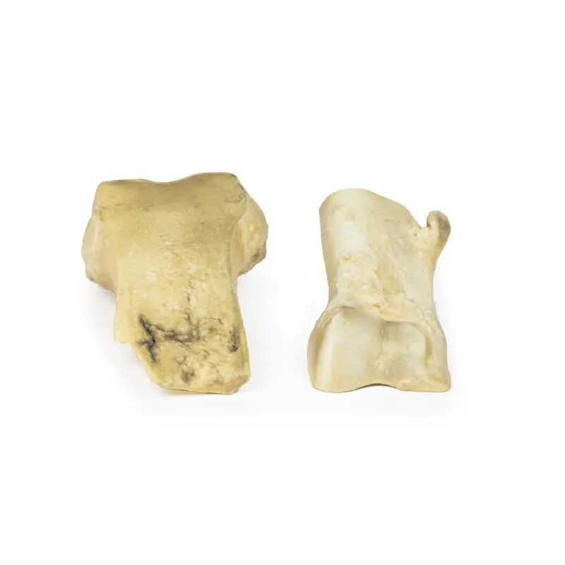
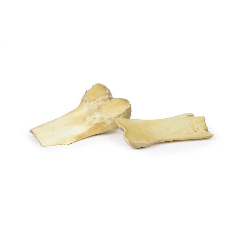
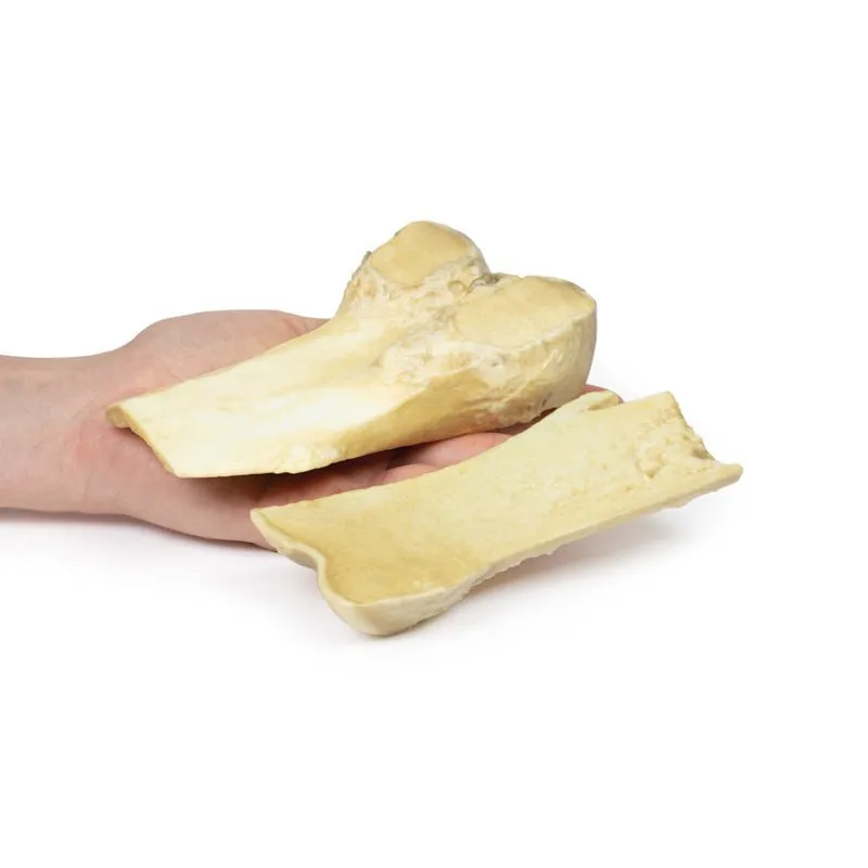
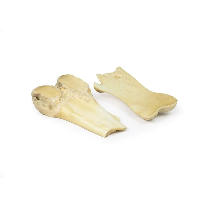
The specimen is the lower end of the patient’s right femur, which has been cut in the
coronal plane and mounted to display the external surfaces. A pedunculated bony protuberance 2 cm in length projects
from the medial aspect of the femoral shaft 7 cm above the medial condyle. The projection is composed of normal bone
with a thin cap of hyaline cartilage at the tip. This is an example of an osteochondroma.
Further Information
An osteochondroma (or an exostosis) is a benign cartilaginous tumour. They
are comprised of a cartilaginous capped bony protrusion from the external surface of the bone from which they arise.
They are the most common benign bone tumours. Most osteochondromas occur spontaneously but they may also occur as
part of multiple hereditary exostosis syndrome or post radiotherapy. They usually develop from or near the growth
plate. They most commonly arise from the appendicular skeleton, especially in the lower limb around the knee or the
upper limb at the proximal humerus. Men are more commonly affected than women.
Symptoms vary on the site and size of the growth. Many osteochondroma remain asymptomatic. Osteochondroma lead to
symptoms from the compression of surrounding neurovascular structures. They may also cause a pain from myositis or a
fracture of the bony spur. They usually present in the second decade of life. They can be diagnosed with plain x-ray
but MRI is the gold standard to ensure that there is no malignancy present within the growth.
Hereditary
exostoses are associated with mutations in the EXT1 and EXT2 genes. Reduced expression of these genes has also been
seen in sporadic osteochondromas. Osteochondromas stop forming as fusion of the growth plate occurs. Treatment of
excision is only if symptoms are severe. Malignant transformation to chondrosarcoma is rare in sporadic cases but
more common in hereditary exostosis (5-20%).
GTSimulators by Global Technologies
Erler Zimmer Authorized Dealer
3D Printed Osteochondroma


")


